
A Comprehensive Guide to Psoriasis Types, and Treatment
Psoriasis is a chronic, non-contagious, immune-mediated, inflammatory skin disease that primarily involves the epidermal cell layer of the skin increasing the cell turnover rate. This produces pruritic, erythematous papules and plaques over the skin, covered by the silvery micaceous scale. The severity of symptoms in psoriasis patients is variable and the clinical course is characterized by recurrent flare-ups and remissions. In addition to its physical manifestations, the disease is also known to have a significant impact on the emotional and psychosocial well-being of the patient. Various topical and systemic agents have been approved for the treatment of psoriasis.

Epidemiology
Psoriasis is one of the most common dermatological diseases, affecting about 2-4 % of the total world’s population. It is found to be more prevalent in countries located more distant from the equator like Europe and Australia compared to those closer to the equator.1 Although it can occur at any age, the incidence of the disease worldwide is higher in adults compared to children. The onset of the disease has a bimodal peak age distribution at < 40 years and > 40 years of age, categorized as Type I psoriasis and Type II psoriasis respectively. More than 75% of the cases have Type I disease. Both men and women are equally affected.
Types of Psoriatic Arthritis
Various types of psoriasis are discussed below, which are mainly classified based on their morphological appearance. The clinical type of psoriasis plays a major role in deciding the treatment protocol.
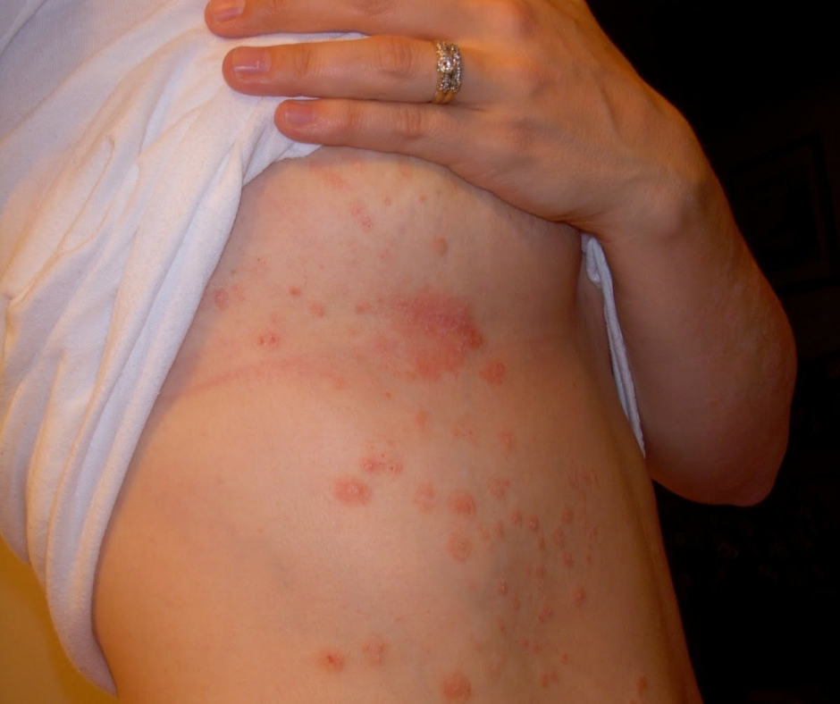
Plaque Psoriasis

It is the commonest type of psoriasis accounting for about 90% of all the affected patients.2 It is also known as psoriasis vulgaris. In plaque-type psoriasis, lesions of variable sizes appear as sharply circumscribed, oval or irregularly shaped, erythematous raised plaques (hence the name plaque psoriasis) which are covered with silvery-white or micaceous scales.
Pruritis is another major symptom expressed by the patients. These lesions are distributed symmetrically and can affect any area of the body, but have a predilection for extensor surfaces of elbows, knees, and back of forearms, scalp, and also the low back area. Psoriatic plaque lesions are further divided into two subphenotypes based on the sizes, which include
- large plaque psoriasis (>3cm) having thick, well-defined, plaques with silvery scales and
- small plaque psoriasis (<3cm) has thin plaques that are either well-defined or ill-defined with fine scales.
Scraping the surface of the psoriatic plaque initially shows silvery scales. Removal of the scales on further scraping reveals bleeding foci on an erythematous background appearing as tiny red pinpoints, the phenomenon of which is known as the” Auspitz sign”. These red pinpoints signify papillomatosis on the tips of dermal papillae.
Psoriatic plaques extending peripherally may acquire different configurations like psoriasis gyrata with predominant curved linear patterns, annular psoriasis with ring-like lesions with central clearing, and psoriasis follicularis with scaly papules at the tips of openings of pilosebaceous follicles.
Whereas healing psoriatic plaque lesions show a white blanching hypopigmented macular ring around them known as the ”Woronff ring“, which is proposed to be related to falling levels of prostaglandins.3
Palmoplantar Psoriasis

This is a localized form of psoriasis with symmetric involvement of both palms and soles. It is frequently characterised by hyperkeratotic plaques with erythema and scaling.
Guttate Psoriasis

The term guttate is derived from the Greek word ”gutta” which translates into ”droplet”. Guttate psoriasis is a rare type accounting for about 2% of all the patients affected with psoriasis. This is commonly seen in children and young adults, presenting 2-4 weeks following an episode of upper respiratory tract infection due to Group B beta-hemolytic streptococci.
Patients can also show elevated titers of antistreptolysin levels. A few cases are also found to be associated with streptococcal perianal dermatitis in children. As the name suggests, lesions in guttate psoriasis appear as acutely developing, multiple, monomorphic, small droplet-like raised lesions or papules with superficial scales.
There can be a generalized distribution of the lesions, but the usual distribution is in a centripetal fashion mainly involving the proximal extremities and trunk. In the majority of cases, guttate psoriasis is a self-limiting disease within 3-4 months. But occasionally it may get complicated and develop into plaque-type psoriasis carrying a bad prognosis.4
Inverse Psoriasis
Occasionally psoriasis may involve the flexor surfaces, which is named inverse or flexural psoriasis. This form is particularly common in obese patients. Lesions occur in the skin creases or intertriginous areas like inguinal folds, perineal region, axillae, and inframammary areas.
Unlike other types of psoriasis, lesions here appear as red, symmetric, well-demarcated fissured plaques without much scaling due to moisture and constant friction in these regions. Inverse psoriasis is relatively resistant to classical treatment regimens.
Erythrodermic Psoriasis

This rare type of psoriasis is the most severe form associated with significant morbidity and mortality risk.5 It is characterized by diffuse erythema of skin involving >80% of the body surface without significant plaques or scaling. Erythrodermic psoriasis usually develops as a complication of chronic plaque-type psoriasis. It can also develop secondary to infections, emotional stress, or sudden withdrawal of drugs like corticosteroids and methotrexate.6 This type of psoriasis is relatively treatment-resistant with a very high risk of fatalities due to cardiac shock. Major complications of erythrodermic psoriasis include:
- Impairment of thermoregulatory capacity of skin because of vasodilation underlying the diffuse erythema, leading to hypothermia.
- Hypoalbuminemia because of protein loss through desquamation, leading to limb edema and high output cardiac failure.
Generalized pustular psoriasis (GPP)

GPP is another rare type of psoriasis and is also known as von Zumbush psoriasis. It is especially common in younger individuals. This type of psoriasis can occur independently or as a complication of psoriasis vulgaris precipitated by the sudden withdrawal of corticosteroids, sun exposure, hypocalcemia, or any other infections.
The familial association has also been identified as another etiological factor, which is largely due to mutations in the IL36RN gene. This results in an increased IL 36 signaling leading to a corresponding increase in proinflammatory cytokines.7 GPP is characterized by the development of sterile, noncontagious pustules on a diffuse erythematous background.
There is also the development of systemic symptoms like fever, chills, malaise, and polyarthralgia associated with increased leukocyte counts. Disease course can be further complicated by frequent relapses, the development of sepsis, and cardiovascular failure.
Management of patients with GPP must be in a hospital setting to ensure adequate hydration and prevent fatalities in disseminated disease.8 There is a pregnancy-associated variant of GPP called impetigo herpetiformis, which is usually seen in the third-trimester pregnancy. The involvement of mucosa and onycholysis are additional features in this variant. There is also and increased risk of fetal anomalies and recurrences during further pregnancies.
Localized palmoplantar pustular psoriasis

This is a localized variant of psoriasis predominantly involving the palms and soles. Onset is frequently between 20 and 60 years of age with a female predilection. Persons with a smoking habit are at particularly high risk. Lesions appear as yellow pustules on an erythematous surface with scaling, which finally gets resolved to leave a dark brown pigment with adherent scaling.
Other Comorbidities
Nail Psoriasis

Nail involvement is a common entity in psoriasis, but psoriatic nail disease poses a diagnostic challenge when presented without cutaneous psoriasis. Psoriatic nail disease has a varied presentation and the commonest of these is the formation of small holes or ”pitting” of the proximal nail matrix due to the loss of parakeratotic cells from the nail plate.
Nail plates can also get dystrophic and ”thickened”. In addition, subungual hyperkeratosis of the nail bed can also result in ”onycholysis”, due to the nail plate getting separated from its nail bed. Onycholysis progresses from the distal part of the nail to the proximal portion. Along with these changes, nails can also get discolored with orangish or brownish-yellow-pigmented areas known as ”oil drop” pigmentation below the nail plate.
Psoriatic Arthritis

Psoriatic arthritis is seronegative arthritis seen in about 30% of psoriatic patients, and in the majority of cases, this develops almost 7-12 years following the onset of skin disease.9 Occasionally arthritis precedes cutaneous involvement or both appear simultaneously. Psoriatic arthritis affects both males and females with equal preponderance. Inflammation of joints mediated by lymphocytes and cytokines like tumor necrosis factor (TNF) play a major role in the pathogenesis of psoriatic arthritis.
Psoriatic patients with nail psoriasis, scalp psoriasis, and perianal psoriasis are at particularly high risk for psoriatic arthritis.10 The vast majority of patients with psoriatic arthritis also develop nail psoriasis. In psoriatic arthritis, both the axial skeleton and peripheral joints are involved. One classical feature is the swelling of entire digits due to dactylitis resulting in the appearance of a ”sausage digit”.
Psoriatic arthritis is clinically diverse and classified based on the criteria developed by Moll and Wright into 5 subtypes:
- Classic type: In this type, distal interphalangeal joints are particularly involved.
- Asymmetric oligoarthritic type: This is the most characteristic type of psoriatic arthritis, in which both major and minor small joints are involved asymmetrically.
- Symmetric polyarthritic type: Symmetrical involvement of multiple large and small joints associated with bony ankylosis.
- Spondylitic type: The majority of these patients have both spondylitis causing back pain and peripheral arthritis.
- Arthritis mutilans: This is named after the clinical appearance of hands following osteolysis of phalangeal and metacarpal bones.
Symmetric polyarthritic type is similar to rheumatoid arthritis (RA), but both of these can be differentiated by the frequent involvement of distal interphalangeal joints and the absence of extra-articular features of RA in psoriatic arthritis. Likewise, the spondylitic type of psoriatic arthritis can be differentiated from ankylosing spondylitis by its less severe course of the disease and better prognosis.
Metabolic Syndrome

Psoriasis has an association with various components of metabolic syndrome like hypertension, diabetes, dyslipidemia causing atherogenesis, and nonalcoholic fatty liver disease. This association is thought to be due to common risk factors shared by both these entities. Metabolic syndrome is particularly common in patients with severe psoriasis.11
Psychiatric Illness

Psoriatic patients are found to be at higher risk for depression and suicidal tendencies. The impact of psoriasis on self-image and quality of life is thought to play a major role in addition to the influence of various cytokines. Young patients and individuals with a severe form of the disease are at particularly high risk compared to others. Psoriatic patients are needed to be screened for depression with tools like Patient Health Questionnaire-2 (PHQ-2).12
Etiopathogenesis of Psoriasis
A complex interplay between genetic, environmental, and immunological factors combined together is involved in the etiopathogenesis of psoriasis.
Genetic Factors

Various family and population-based studies confirm the genetic basis for the development of psoriasis. A relatively higher risk of psoriasis is observed in monozygotic twins compared to dizygotic twins, but this concordance rate in monozygotic twins is not always complete. This suggests the additional influence of environmental factors in individuals who are genetically predisposed to psoriasis.13
The inheritance pattern in susceptible individuals was previously thought to be an autosomal dominant pattern, but now it is proven to be a multifactorial pattern of inheritance.14 Various genes involved in the pathogenesis of psoriasis are mapped by different methods like linkage analysis and genome-wide association studies. Linkage analysis studies resulted in the detection of major determinant loci of psoriasis called ”psoriasis susceptibility 1” or PSORS1, within the major histocompatibility complex (MHC) region on chromosome 6p21.3.
The majority of psoriasis-associated alleles are isolated from the MHC region. Human Leukocyte Antigen Cw* 0602 (HLA-Cw* 0602) is one such allele that is found to have a strong association with type 1 psoriasis (early-onset before 40 years) and is also found in almost all the patients with guttate psoriasis. HLA-C has the capacity to regulate both innate and adaptive immune responses but the exact mechanism of how this HLA-C region predisposes to psoriasis is still not explained clearly. Other common HLA alleles found in psoriatic patients are HLA B57, HLA B17, and HLA B47.
Several genome-wide association studies and a meta-analysis by Tsoi et al.,15 identified a number of other psoriasis susceptibility genes. A susceptibility locus associated with a dominant pattern of inheritance was identified at chromosome 17q25, known as PSORS2. Among the psoriatic susceptibility genes identified; LCE, KLF4, and ETS1 are found to be skin-specific which plays a key role in skin barrier formation. Other important genes involved belong to the NF-kB pathway of innate immunity which regulates the activation of NF-kB downstream from TNF and IL-17 and thus mediates the chronic inflammatory process. Genes belonging to the IL-23/IL-17 pathway also play a role in the pathogenesis of psoriasis.16 A few of these genes are also responsible for the development of other immune-mediated conditions like Crohn’s disease in these patients.
Environmental factors

Environmental factors involved in triggering the psoriasis disease process include drugs, infections, stress, UV exposure, smoking, alcohol, diet, and obesity; and the majority of these are potentially modifiable risk factors. Drugs like lithium, beta-blockers, imiquimod, interferons, and anti-TNF antibodies are associated with the aggravation of the disease.
Imiquimod, a toll-like receptor 7/8 agonist used in the treatment of genital warts is the most extensively studied drug in this respect, which acts as a trigger of psoriasis through the activation of the Interferon signaling pathway. Infections that are commonly associated with psoriasis are Human Immunodeficiency Virus and Streptococcus.
Obesity is another important risk factor for psoriasis. Most studies theorize this association to be due to an association between adipocytes and inflammatory-type macrophages. Obesity also affects the efficacy of various drugs used in the management of psoriasis by interfering with their pharmacokinetics.
Trauma to the skin can also trigger psoriatic changes at the injured site, the phenomenon of which is labeled as the ”koebner phenomenon”.
Immune factors

Both innate and adaptive immune responses are involved in the pathogenesis of psoriasis. Environmental triggers trigger the initiation of a dysregulated immune response in a genetically predisposed individual. T cells are critical cells involved in this process, which are activated by antigen-presenting cells like Langerhans cells.
In addition, cytokines like IL-12, 23 secreted by these antigen-presenting cells mediate the differentiation of T cells into Th1 and Th17 cells. These activated T cells are further stimulated to produce various cytokines like IFN-gamma and IL-17, that are involved in the pathogenic changes that occur in the skin of psoriatic patients.
Mechanism of Psoriasis
Environmental and genetic factors combined together result in a dysregulated immune response that triggers an inflammatory process in the skin. Keratinocytes (KC) are the major skin cells to be affected in this process. In normal healthy skin, keratinocytes found in the outermost epidermal layer of skin are shed within four weeks after passing through various stages of development.
In psoriatic patients, these cells hyperproliferate and undergo rapid cell turnover resulting in quick shedding within a few days. Hyperproliferative keratinocytes result in the formation of thick skin (acanthosis) and loss of the granular layer of the epidermis with keratinocytes retaining their nuclei in the cornified layer resulting in scaling of the skin (parakeratosis).17
How to Diagnose Psoriasis?
Diagnosis of psoriasis is based primarily on its characteristic clinical features like the presence of well-defined, itchy, erythematous plaques with silvery scales (plaque-type psoriasis). There can also be other less common types of lesions seen in various other variants of psoriasis or even nail involvement or joint involvement.

A biopsy of skin lesions or skin scraping may be needed when the diagnosis is in doubt to differentiate from other dermatological conditions. Histopathological examination of the skin section will show epidermal thickening or acanthosis, which is the characteristic feature. Other features are parakeratosis, loss of stratum granulosum layer of the epidermis, kogoj spongiotic pustules, and Munro microabscesses.18
In addition to this, a large number of activated inflammatory T cells can be seen, of which CD8+ T cells are predominant in the epidermal layer and CD4+ T cells in the dermal layer of the skin.19 Skin biopsy is also helpful to rule out any suspected fungal infections. In nail psoriasis, a nail sample can also be tested to rule out fungal nail infection.

Laboratory investigations like rheumatic factor and uric acid levels can be done to differentiate psoriatic arthritis from rheumatoid arthritis and gouty arthritis respectively. However uric acid levels may sometimes be elevated even in the pustular type of psoriasis. Radiographs of the involved joints may also be necessary for certain situations to differentiate from psoriatic arthritis.
After making a diagnosis of psoriasis, it is essential to grade the disease severity which helps in guiding the treatment protocol. In general, if greater than 10% of the body surface area is affected, regions like the face, hands, feet, and genitalia are involved; and quality of life is affected, psoriasis is considered to be severe.
The severity of psoriasis can also be graded with the help of measuring tools like the dermatology life quality index (DLQI) score or the psoriasis area severity index (PASI). PASI (ranging from 0 to 72) is the most commonly used method and it can also be used to assess the effectiveness of the treatment initiated. PASI assesses psoriasis based on four factors which include the degree of redness of the skin, amount of skin shedding, plaque thickness, and size of the surface area of the skin affected.
Differential Diagnosis
Common dermatological conditions similar to the appearance of psoriasis are discoid eczema, lichen planus, atopic and contact dermatitis, tinea corporis, and pityriasis rosea. Lichen planus has violaceous lesions and mucosa is also involved. Pityriasis rosea which may be confused with guttate psoriasis has pink oval papules or patches and; the face and distal extremities are usually spared. Fungal infections can be differentiated by testing for a skin biopsy. Occasionally secondary syphilitic rash can be confused with psoriasis, but the lessons here are usually copper-colored with frequent palms and soles involvement.
Nail psoriasis can be differentiated from similar-looking fungal nail infections by examining nail specimens. Rheumatoid arthritis and gouty arthritis are important differential diagnoses for psoriatic arthritis both of which can be differentiated based on laboratory investigations and radiography.
Psoriasis Treatment
Psoriasis is a disease with no definitive cure and all the available treatment strategies are targeted at reducing the disease activity. Before the initiation of therapy, the severity of the disease is assessed using PASI and; graded into mild, moderate, and severe categories. Basic skincare with emollients to relieve dryness and itching is required in all patients. Whenever identified, any environmental factors triggering the episodes of psoriasis must be treated accordingly.
Topical Agents

Topical agents are the first drugs of choice in mild to moderate psoriasis. Corticosteroids and vitamin D analogues are commonly used, topical agents. Topical corticosteroids which act as an anti-inflammatory are generally very effective in mild to moderate psoriasis and also well tolerated. They can also be given in combination with keratolytic agents like salicylic acid.
Other topical agents are vitamin D analogues, which act inhibition of T cell activity and also inhibition of keratinocyte proliferation. These are non-inferior to topical steroids in efficacy and are considered relatively safer compared to topical corticosteroids for long-term usage.
Common side effect seen with these agents is irritant dermatitis and very occasionally hypercalcemia. Vitamin D analogues are found to be better tolerated and more efficacious when given in combination with topical steroids like betamethasone.20
Systemic Therapy

Systemic therapies are indicated in moderate to severe disease or mild disease unresponsive to first-line topical agents. Phototherapy or light therapy using ultraviolet light (UV) is one of the common most systemic therapies indicated in the management of psoriasis. Both UVA and UVB can be used, of which narrowband UVB (NB-UVB) is often used as first-line therapy.
NB-UVB induces DNA damage and interferes with the progression of the cell cycle targeting the rapid cell turnover rate seen in psoriasis. This therapy is generally well tolerated except for side effects like redness, and itching of the skin. The only limitation of UVB therapy is the limited availability of phototherapy centers.
Another type of phototherapy commonly used is PUVA therapy, in which a photosensitizer called psoralen is ingested by the patient prior to phototherapy with UVA. Psoralen gets activated on exposure to UV light and suppresses the immune mechanisms in the skin of patients with psoriasis.
Though PUVA therapy is more efficacious compared to UVB therapy, a major limitation is its association with an increased risk of squamous cell carcinoma after prolonged therapy.21 Both PUVA and UVB therapies are usually given in combination with other topical or systemic agents.
Acitretin, a synthetic retinoid is another systemic agent used in the treatment of moderate to severe psoriasis. It is almost always used in combination therapy with other agents but never as monotherapy. Usual side effects include dryness of the skin and gastrointestinal upset. This is also contraindicated in women of childbearing age because of its potential teratogenicity.

Other immunosuppressant systemic agents commonly used are methotrexate and cyclosporine which are commonly associated with side effects like marrow, hepatic toxicity (methotrexate), and renal toxicity (cyclosporine).
Methotrexate is a folate antagonist, the action of which is mediated through its immunosuppressant properties. The major limitation of this drug is its hepatotoxicity.
Cyclosporine is a calcineurin inhibitor that in particular has shown positive efficacy results in patients with psoriatic arthritis. Major side effects include nephrotoxicity and electrolyte disturbances. Both of these drugs are not preferable for pregnant women.
Newer Biologic Agents
Several biologic agents have recently emerged which can be tried in patients who are refractory to traditional systemic therapeutic agents. In contrast to immunosuppressant drugs discussed earlier, biological agents target specific components of the immune system. However,
the major limitation of all these drugs is their association with an increased risk of infections. The first biological agents to be approved for the treatment of psoriasis are anti-T-cell agents- Alefacept and Efalizumab.
Alefacept acts by binding to CD2 on T cells and then inducing cellular apoptosis. A study by Kruger et al. has shown positive results with about 40% of patients starting on Alefacept achieving a 75 % reduction in PASI.22
Efalizumab which acts by inhibiting T-cell migration has recently been withdrawn from the market due to three reported cases of progressive multifocal leukoencephalopathy.23
Another group of biologic agents is anti-cytokine agents targeting cytokine TNF, which plays a critical role in the pathogenesis of psoriasis. Anti-TNF antibodies like Adalimumab and Infliximab are the commonly used drugs in this category with studies showing about 75% improvement in the PASI of patients.24, 25
Etanercept, which is a TNF receptor fusion protein also showed similar results with significant improvement in PASI.26 Other anti-TNF agents are certolizumab and golimumab which have also been approved for the treatment of psoriatic arthritis.
In addition to the increased risk of infections, there has also been an increased risk of non-melanoma skin cancer with anti-TNF agents.
Another more recent biologic agent approved is ustekinumab, which targets the p40 subunit on both IL-23/12 and thus inhibits the initiation of T helper cell responses. Three newer biologics Brodalumab, Ixekizumab, and Secukinumab; targeting cytokine IL-17 are still under trial but data available till now has shown positive results.
Though proven to be efficacious, biologic agents do have certain limitations like loss of responsiveness due to the development of anti-drug antibodies and increased risk of infections.
Novel Small Molecule Drugs
To overcome the limitations of biological agents and also to reduce the economic burden caused by biological agents, novel treatment options like low molecule drugs are now being considered. The most widely known drug in this category is Tofacitinib, which is a Janus kinase inhibitor. However, these drugs are under trial and still not approved for the treatment of psoriasis.
Conclusion
Recent understanding of the etiopathogenesis of psoriasis in detail has led to the development of various newer treatment strategies, the majority of them with positive efficacy results. However, psoriasis still remains a disease that is not completely curable and there is a need for exploration of additional definitive treatment strategies with a better safety profile.
References
1. Parisi, R., Symmons, D., Griffiths, C., u0026amp; Ashcroft, D. (2013). Global Epidemiology of Psoriasis: A Systematic Review of Incidence and Prevalence. Journal Of Investigative Dermatology, 133(2), 377-385. doi: 10.1038/jid.2012.339.
2. Boehncke, W., u0026amp; Schön, M. (2015). Psoriasis. The Lancet, 386(9997), 983-994. doi: 10.1016/s0140-6736(14)61909-7.
3. Van DE Kerkhof. (1998). Correspondence. The Woronoff zone surrounding the psoriatic plaque. British Journal Of Dermatology, 139(1), 167-168. doi: 10.1046/j.1365-2133.1998.02347.x.
4. Martin, B. (1996). How Great Is the Risk of Further Psoriasis Following a Single Episode of Acute Guttate Psoriasis?. Archives Of Dermatology, 132(6), 717. doi: 10.1001/archderm.1996.03890300147032.
5. Gudjonsson JE, Elder JT. Psoriasis. Fitzpatrick’s Dermatology in General Medicine. New York: McGraw Hill; 2008. pp. 169–94.
6. Ayala F 2007. Clinical presentation of psoriasis. Reumatismo 59: 40–45.
7. Onoufriadis A, Simpson MA, Pink AE, Di Meglio P, Smith CH, Pullabhatla V, Knight J, Spain SL, Nestle FO, Burden AD, et al. 2011. Mutations in IL36RN/IL1F5 are associated with the severe episodic inflammatory skin disease known as generalized pustular psoriasis. Am J Hum Genet 89: 432–437.
8. Gülekon A. Psöriasis ve benzeri dermatozlar. In: Tüzün Y, Gürer MA, Serveroğlu S, Sungur O, Aksungur LA, editors. Dermatoloji. 3. baskı. İstanbul: Nobel Tıp; 2008. pp. 745–60.
9. Mease PJ, Armstrong AW. Managing patients with psoriatic disease: the diagnosis and pharmacologic treatment of psoriatic arthritis in patients with psoriasis. Drugs 2014;74(4):423-41.
10. Wilson FC, Icen M, Crowson CS, McEvoy MT, Gabriel SE, Kremers HM. Incidence and clinical predictors of psoriatic arthritis in patients with psoriasis: a population-based study. Arthritis Rheum 2009;61(2):233-9. Erratum in: Arthritis Rheum 2010;62(4):574.
11. Gisondi, P., Fostini, A., Fossà, I., Girolomoni, G., u0026amp; Targher, G. (2018). Psoriasis and the metabolic syndrome. Clinics In Dermatology, 36(1), 21-28. doi: 10.1016/j.clindermatol.2017.09.005.
12. Korman, A., Hill, D., Alikhan, A., u0026amp; Feldman, S. (2016). Impact and management of depression in psoriasis patients. Expert Opinion On Pharmacotherapy, 17(2), 147-152. doi: 10.1517/14656566.2016.1128894.
13. Elder, J., Bruce, A., Gudjonsson, J., Johnston, A., Stuart, P., u0026amp; Tejasvi, T. et al. (2010). Molecular Dissection of Psoriasis: Integrating Genetics and Biology. Journal Of Investigative Dermatology, 130(5), 1213-1226. doi: 10.1038/jid.2009.319.
14. Elder JT, Nair RP, Voorhees JJ. Epidemiology and genetics of psoriasis. J Invest Dermatol. 1994;102:24S–28S.
15. Tsoi LC, Spain SL, Knight J, Ellinghaus E, Stuart PE, Capon F, Ding J, Li Y, Tejasvi T, Gudjonsson JE, et al. 2012. Identification of 15 new psoriasis susceptibility loci highlights the role of innate immunity. Nat Genet 44: 1341–1348.
16. Di Cesare A, Di Meglio P, Nestle FO 2009. The IL-23/Th17 axis in the immunopathogenesis of psoriasis. J Invest Dermatol 129: 1339–1350.
17. Bhalerao J, Bowcock AM. The genetics of psoriasis: a complex disorder of the skin and immune system. Hum Mol Genet. 1998;7:1537–45.
18. Elston DM, Ferringer T, Ko C, Peckham S, High W, DiCaudo D. Dermatopathology. 2nd ed. Philadelphia, Pa: Elsevier Saunders; 2013.
19. Raychaudhuri, S., Maverakis, E., u0026amp; Raychaudhuri, S. (2014). Diagnosis and classification of psoriasis. Autoimmunity Reviews, 13(4-5), 490-495. doi: 10.1016/j.autrev.2014.01.008.
20. Schlager, J., Rosumeck, S., Werner, R., Jacobs, A., Schmitt, J., Schlager, C., u0026amp; Nast, A. (2016). Topical treatments for scalp psoriasis: summary of a Cochrane Systematic Review. British Journal Of Dermatology, 176(3), 604-614. doi: 10.1111/bjd.14811.
21. Menter A, Korman NJ, Elmets CA, Feldman SR, Gelfand JM, Gordon KB, et al. Guidelines of care for the management of psoriasis and psoriatic arthritis: Section 5. Guidelines of care for the treatment of psoriasis with phototherapy and photochemotherapy. J Am Acad Dermatol. 2010 Jan. 62(1):114-35.
22. Krueger GG, Papp KA, Stough DB, Loven KH, Gulliver WP, Ellis CN 2002. A randomized, double-blind, placebo-controlled phase III study evaluating efficacy and tolerability of 2 courses of alefacept in patients with chronic plaque psoriasis. J Am Acad Dermatol 47: 821–833.
23. Tan CS, Koralnik IJ 2010. Progressive multifocal leukoencephalopathy and other disorders caused by JC virus: Clinical features and pathogenesis. Lancet Neurol 9: 425–437.
24. Chaudhari U, Romano P, Mulcahy LD, Dooley LT, Baker DG, Gottlieb AB. Efficacy and safety of infliximab monotherapy for plaque-type psoriasis: a randomised trial. Lancet. 2001;357:1842–7. doi: 10.1016/S0140-6736(00)04954-0.
25. Menter A, Tyring SK, Gordon K, Kimball AB, Leonardi CL, Langley RG, Strober BE, Kaul M, Gu Y, Okun M, Papp K. Adalimumab therapy for moderate to severe psoriasis: A randomized, controlled phase III trial. J Am Acad Dermatol. 2008;58:106–15. doi: 10.1016/j.jaad.2007.09.010.
26. Tyring S, Gottlieb A, Papp K, Gordon K, Leonardi C, Wang A, Lalla D, Woolley M, Jahreis A, Zitnik R, et al. 2006. Etanercept and clinical outcomes, fatigue, and depression in psoriasis: Double-blind placebo-controlled randomised phase III trial. Lancet 367: 29–35.
27. Ports WC, Khan S, Lan S, Lamba M, Bolduc C, Bissonnette R, Papp K 2013. A randomized phase 2a efficacy and safety trial of the topical Janus kinase inhibitor tofacitinib in the treatment of chronic plaque psoriasis. Br J Dermatol 169: 137–145.